PD-1 (Woodchuck) / NFAT - Reporter - Jurkat Recombinant Cell Line
Catalog #
79456
$10,340
*
●
●
Purchase
Description
Recombinant Jurkat T cell expressing firefly luciferase gene under the control of NFAT response elements with constitutive expression of woodchuck (groundhog, Marmota monax) PD-1 (Programmed Cell Death 1, PDCD1, SLEB2, CD279, GenBank Accession #HQ403652).

Interested in screening and profiling inhibitors, blocking antibodies, or activators of woodchuck PD-1 without the need to purchase and license the cell line? Check out our Cell Signaling Pathway Screening.
Purchase of this cell line is for research purposes only; commercial use requires a separate license. View the full terms and conditions.
●
Synonyms
Groundhog, Marmota Monax, PD-1, Programmed Cell Death 1, PDCD1, SLEB2, CD279, NFAT
●
Product Data Gallery
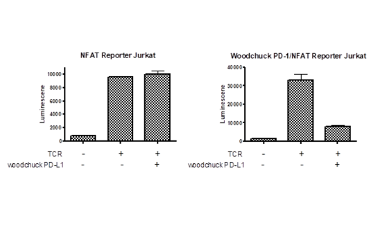
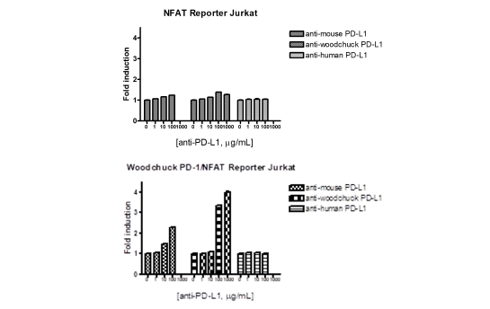
The reporter activity from woodchuck PD-1/NFAT reporter Jurkat cells is decreased when co-cultured with HEK293 cells transiently transfected with woodchuck PD-L1
Woodchuck PD-1/PD-L1 cell-based assay using the woodchuck PD-1/NFAT Reporter-Jurkat cells and woodchuck PD-L1/ TCR Activator CHO cells
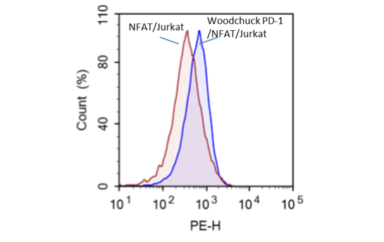
FACS analysis of cell surface expression of woodchuck PD-1 in PD-1/NFAT Reporter-Jurkat cells
Product Info
Storage and Usage
Citations
Host Cell Line
Jurkat
Supplied As
Each vial contains 2 x 10^6 cells in 1 ml of 10% DMSO
Materials Required But Not Supplied
- • HEK293 Cell and its Growth Medium
- • TCR Activator/Woodchuck PD-L1 Mammalian Expression Kit (BPS Bioscience #79455)
- • Transfection reagent for mammalian cell line [We use Lipofectamine™ 2000 (Life technologies #11668027). However, other transfection reagents work equally well.]
- • Opti-MEM I Reduced Serum Medium (life technologies #31985-062)
- • Thaw Medium 2 (BPS Bioscience #60184)
- • Growth Medium 2A (BPS Bioscience #60190)
- • Assay Medium: Thaw Medium 2 (BPS Bioscience #60184)
- • 96-well tissue culture treated white clear-bottom assay plate (Corning, #3610)
- • ONE-Step™ Luciferase Assay System (BPS Bioscience, #60690)
- • Luminometer
Mycoplasma Testing
The cell line has been screened using the PCR-based Venor®GeM Mycoplasma Detection kit (Sigma-Aldrich, #MP0025) to confirm the absence of Mycoplasma species.
Background
The binding of Programmed Cell Death Protein 1 (PD-1), a receptor expressed on activated Tcells, to its ligands, PD-L1 and PD-L2, negatively regulates immune responses. The PD-1 ligands are found on most cancers, and PD-1:PD-L1/2 interaction inhibits T cell activity and allows cancer cells to escape immune surveillance. The PD-1:PD-L1/2 pathway is also involved in regulating autoimmune responses, making these proteins promising therapeutic targets for a number of cancers, as well as multiple sclerosis, arthritis, lupus, and type I diabetes.
 Powered by Bioz
6405 Mira Mesa Blvd. Suite 100
San Diego, CA 92121
858-202-1401
San Diego, CA 92121
858-202-1401
Products are for Research Use Only
COMPANY
Copyright © 2024
BPS Bioscience, Inc.
All rights reserved.
Diagrams on this website are created with BioRender.com.