PD-1/IL-2 Luciferase Reporter Jurkat Cell Line
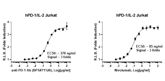
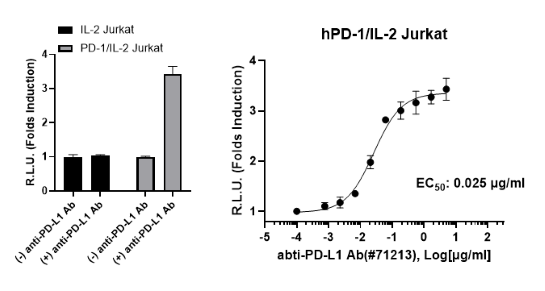
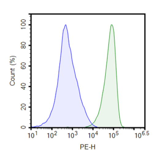
PD-1/IL-2 Luciferase Reporter Jurkat Cell Line is a Jurkat T cell line constitutively expressing human PD-1 (Programmed Cell Death 1, also known as PDCD1, SLEB2, CD279, GenBank Accession #NM_005018), and conditionally expressing firefly luciferase under the control of a human interleukin-2 (IL-2) promoter. This cell line has been validated by flow cytometry for expression of PD-1, and in co-culture assays that assess the PD-L1/PD-1 interaction on Jurkat T cell activation.
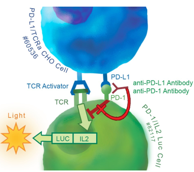
Figure 1: Illustration of the mechanism of action of PD-1/IL-2 Luciferase Reporter Jurkat Cell Line in a co-culture assay.
The TCR activator presented at the surface of PD-L1/TCR Activator CHO cells stimulates TCR (T cell receptor) in Jurkat T cells, whereas overexpression of PD-L1 on the CHO cell line engages Jurkat PD-1 resulting in blocking TCR signaling and preventing activation of the IL-2 promoter. Addition of a neutralizing anti-PD-1 or anti-PD-L1 antibody to the co-culture prevents formation of the PD-L1/PD-1 complex, and results in TCR activation and increased IL-2 promoter activity, which translates into increased luciferase reporter signal.
Interested in screening and profiling inhibitors, blocking antibodies, or activators of PD-1 without the need to purchase and license the cell line? Check out our Cell Signaling Pathway Screening.
Purchase of this cell line is for research purposes only; commercial use requires a separate license. View the full terms and conditions.
Media Required for Cell Culture
| Name | Ordering Information |
| Thaw Medium 2 | BPS Bioscience #60184 |
| Growth Medium 2A | BPS Bioscience #60190 |
Materials Required for Cellular Assay
| Name | Ordering Information |
| PD-L1/TCR Activator CHO Cell Line | BPS Bioscience #60536 |
| IL-2 Luciferase Reporter Jurkat Cell Line | BPS Bioscience #60481 |
| Thaw Medium 3 | BPS Bioscience #60186 |
| Anti-PD-1 Neutralizing Antibody | BPS Bioscience #71120 |
| Anti-PD-L1 (CD274) Neutralizing Antibody | BPS Bioscience #71213 |
| Anti-CD28 Agonist Antibody (Humanized) | BPS Bioscience #100186 |
| Nivolumab | Selleckchem #A2002 |
| ONE-Step™ Luciferase Assay System | BPS Bioscience #60690 |
| 96-well tissue culture-treated white clear-bottom assay plate | |
| Luminometer |
The cell line has been screened to confirm the absence of Mycoplasma species.
PD-L1 and PD-L2 binding to PD-1, a receptor expressed on T cells, negatively regulates immune responses. PD-1 ligands PD-L1 and PD-L2 are found on the surface of most cancer cells, and their interaction with receptor PD-1 inhibits T cell activity and allows cancer cells to escape immune surveillance. This pathway is also involved in regulating autoimmune responses. Therefore, these proteins (termed immune checkpoints) are promising therapeutic targets for many types of cancer as well as multiple sclerosis, arthritis, lupus, and type I diabetes. Checkpoint inhibitors have remarkable efficacy in a wide range of cancer types and have revolutionized cancer treatment. PD-1 inhibitors nivolumab, pembrolizumab, cemiplimab and PD-L1 inhibitors atezolizumab, avelumab, and durvalumab are all FDA-approved drugs for immuno-therapy.
Sasca D, et al. 2019 Blood 133: 2305-2319
 Powered by Bioz
San Diego, CA 92121
858-202-1401
Products are for Research Use Only
COMPANY
Copyright © 2024
BPS Bioscience, Inc.
All rights reserved.
Diagrams on this website are created with BioRender.com.