LAG3 / NFAT Reporter - Jurkat Recombinant Cell Line
Catalog #
71278
$11,095
*
●
●
Purchase
Description
Recombinant Jurkat T cell expressing firefly luciferase gene under the control of NFAT response elements with constitutive expression of human LAG3 (lymphocyte-activation gene 3, CD223, GenBank Accession # NM_002286). 
Interested in screening and profiling inhibitors, blocking antibodies, or activators of LAG3 without the need to purchase and license the cell line? Check out our Cell Signaling Pathway Screening.
Purchase of this cell line is for research purposes only; commercial use requires a separate license. View the full terms and conditions.
●
Synonyms
LAG3, LAG-3, CD223, Cell Line, Jurkat
●
Product Data Gallery
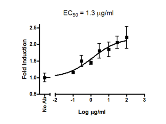
Characterization of biological activity of anti-LAG3 neutralizing antibody in LAG3 cell-based assay using the LAG3/NFAT Reporter-Jurkat cells co-cultured with Raji cells.
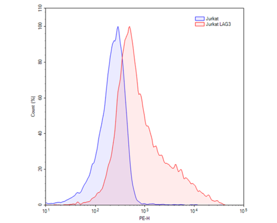
FACS analysis of cell surface expression of LAG3 in LAG3/NFAT Reporter-Jurkat cells
Product Info
Storage and Usage
Citations
Materials Required But Not Supplied
- Raji cell (ATCC # CCL-86)
- Assay medium: RPMI1640 + 10% FBS + 1% Penicillin/Streptomycin (Thaw Medium 2 BPS Bioscience #60184)
- Growth Medium 2A (BPS Cat. #60190)
- Small glass vials
- Tissue culture grade water
- Superantigen (SEE, Toxin Technology # ET404), (stock = 1mg/ul, prepared in tissue culture grade water, in a glass vial and stored at-20°C, minimize freeze/thaw cycles).
- Anti-LAG-3 neutralizing antibody (BPS Cat. #71219)
- 96-well tissue culture-treated white clear-bottom assay plate
- ONE-Step luciferase assay system (BPS Cat. # 60690) or other luciferase reagents for measuring firefly luciferase activity
- Luminometer
Format
Each vial contains 2 x 10^6 cells in 1 ml of 10% DMSO
UniProt #
P18627
Mycoplasma Testing
The cell line has been screened using the PCR-based Venor®GeM Mycoplasma Detection kit (Sigma-Aldrich) to confirm the absence of Mycoplasma species.
Background
Lymphocyte-activation gene 3 (LAG3, CD223) is a cell surface protein that belongs to immunoglobulin (Ig) superfamily. LAG3 is expressed on activated T cells, natural killer cells, B cells, and plasmacytoid dendritic cells. Its main ligand is MHC class II, to which it binds with higher affinity than CD4. It negatively regulates cellular proliferation, activation, and homeostasis of T cells, in a similar fashion to CTLA-4 and PD-1, and has been reported to play a role in Treg suppressive function. A number of LAG3 antibodies are in preclinical development for treatments for cancer and autoimmune disorders. LAG3 may be a better immune checkpoint inhibitor target than CTLA-4 or PD-1 since antibodies to these two checkpoints are only activating effector T cells, and not inhibiting Treg activity where an antagonist LAG3 antibody can both activate effector T cells (by downregulating the LAG3 inhibiting signal) and inhibit induced (i.e. antigen-specific) Treg suppressive activity.
 Powered by Bioz
6405 Mira Mesa Blvd. Suite 100
San Diego, CA 92121
858-202-1401
San Diego, CA 92121
858-202-1401
Products are for Research Use Only
COMPANY
Copyright © 2024
BPS Bioscience, Inc.
All rights reserved.
Diagrams on this website are created with BioRender.com.