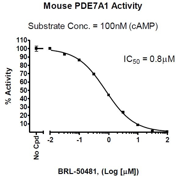
The Mouse PDE7A Assay Kit is designed for identification of inhibitors of Mouse PDE7A using fluorescence polarization. The assay is based on the binding of a fluorescent nucleotide monophosphate generated by Mouse PDE7A to the binding agent.
The key to the Mouse PDE7A Assay Kit is the specific binding agent. Using this kit, only two simple steps on a microtiter plate are required for Mouse PDE7A reactions. First, the fluorescently labeled cAMP is incubated with a sample containing Mouse PDE7A for 1 hour. Second, a binding agent is added to the reaction mix to produce a change in fluorescent polarization that can then be measured using a fluorescence reader equipped for the measurement of fluorescence polarization.
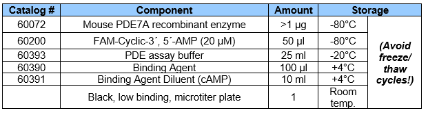
The Mouse PDE7A Assay Kit comes in a convenient 96-well format, with purified Mouse PDE7A enzyme, fluorescently labeled substrate (cAMP), binding agent, and PDE assay buffer for 100 enzyme reactions.
Materials Required But Not Supplied
Fluorescent microplate reader capable to measure fluorescence polarization.
Adjustable micropipettor and sterile tips.
1,4-Dithiothreitol (DTT) 1 M in anhydrous DMSO.
Format
COMPONENTS:
Background
Phosphodiesterases (PDEs) play an important role in the dynamic
regulation of cAMP and cGMP signaling. PDE10A is a dual substrate PDE highly
expressed in striatal medium spiny neurons. PDE10A inhibitors can improve the
cognitive symptoms of schizophrenia, and exhibit potential therapeutic value for
Huntington’s Disease. Phosphodiesterases catalyze the hydrolysis of the phosphodiester bond in dye-labeled
cyclic monophosphates. Beads selectively bind the phosphate group in the nucleotide
product. This increases the size of the nucleotide relative to unreacted cyclic
monophosphate. In the polarization assay, dye molecules with absorption transition
vectors parallel to the linearly-polarized excitation light are selectively excited. Dyes
attached to the rapidly-rotating cyclic monophosphates will obtain random orientations
and emit light with low polarization. Dyes attached to the slowly-rotating nucleotide-bead
complexes will not have time to reorient and therefore will emit highly polarized light.
References
1. Yang, G., McIntyre, K.W., Townsend, R.M.,et al., Phosphodiesterase 7A-Deficient Mice Have Functional T Cells, J. Immunology December 15, 2003, 171 (12):6414-6420.
2. Lee, R., Wolda, S., Moon, E., Esselstyn, J., Hertel, C., Lerner, A. PDE7A is expressed in human B-lymphocytes and is up-regulated by elevation of intracellular cAMP, Cellular Signalling, 2002, 14(3): 277-284.
Storage/Stability
At least 6 months from date of receipt, when stored as directed. Kit components require different storage conditions. Be sure to store each component at the proper temperature upon arrival.
Instructions for Use
See assay kit data sheet for detailed protocol.
Shipping Temperature
-80°C
Contraindications
DMSO >1%, strong acids or bases, ionic detergents, high salt
Warnings
This assay requires a fluorescent microplate reader capable of measuring fluorescence polarization (FP) and equipped with the required partsto read the FP signal. For more information FP technology, visit our Tech Note: FP, assay principles and applications.
Avoid freeze/thaw cycles
Disclaimers and Limitations
BPS Bioscience assay kits are validated using the listed components according to our specific protocol. Any deviations to kit components or protocols, such as using different proteins, cell/tissue lysates, or buffers (including using the same component from other commercial sources) will void any warranties on the performance of the assay kit and is not recommended.
End-users should immediately report any issues to [email protected] upon receipt of the kit, such as missing components, damaged vials, no dry ice, etc.
 Powered by Bioz