NF-κB (GFP) – Reporter HEK293 Recombinant Cell Line
Catalog #
79402
$2,340*
●
Purchase
●
Description
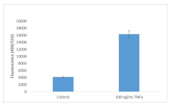
The NF-κB reporter (GFP)-HEK293 cell line is designed for monitoring the nuclear factor Kappa B (NF-κB) signal transduction pathways. It contains the gene for green fluorescent protein (GFP) driven by four copies of NF-κB response element located upstream of the minimal TATA promoter. After activation by pro-inflammatory cytokines or stimulants of lymphokine receptors, endogenous NF-κB transcription factors bind to the DNA response elements, inducing transcription of the GFP gene.
Interested in screening and profiling inhibitors, blocking antibodies, or activators of NF-κB-mediated signaling without the need to purchase and license the cell line? Check out our Cell Signaling Pathway Screening.
Purchase of this cell line is for research purposes only; commercial use requires a separate license. View the full terms and conditions.
This cell line has been screened using the Venor™ GeM Mycoplasma Detection Kit, PCR
Based (Sigma, #MP0025) to confirm the absence of Mycoplasma contamination.
Background
Nuclear factor-κB (NF-κB)/Rel proteins include NF-κB2 p52/p100, NF-κB1 p50/p105, c-Rel, RelA/p65, and RelB. These proteins function as dimeric transcription factors that control genes regulating a broad range of biological processes including innate and adaptive immunity, inflammation, stress responses, B cell development, and lymphoid organogenesis. In the classical (or canonical) pathway, NF-κB/Rel proteins are bound and inhibited by IκB proteins. Proinflammatory cytokines, LPS, growth factors, and antigen receptors activate an IKK complex (IKKβ, IKKα, and NEMO), which phosphorylates IκB proteins. Phosphorylation of IκB leads to its ubiquitination and proteasomal degradation, freeing NF-κB/Rel complexes. Active NF-κB/Rel complexes are further activated by phosphorylation and translocated to the nucleus where they induce target gene expression. In the alternative (or noncanonical) NF-κB pathway, NF-κB2 p100/RelB complexes are inactive in the cytoplasm. Signaling through a subset of receptors, including LTβR, CD40, and BR3, activates the kinase NIK, which in turn activates IKKα complexes that phosphorylate C-terminal residues in NF-κB2 p100. Phosphorylation of NF-κB2 p100 leads to its ubiquitination and proteasomal processing to NF-κB2 p52, creating transcriptionally competent NF-κB p52/RelB complexes that translocate to the nucleus and induce target gene expression.
References
1. Pessara U, Koch N (1990). Mol. Cell Biol.10(8):4146-4154.
2. Baeuerle PA (1998). Curr. Biol. 8(1):R19-R22.
3. Takada Y, Kobayashi Y, Aggarwal BB (2005). J. Biol. Chem. 280(17):17203-17212.
Storage/Stability
Store in liquid nitrogen immediately upon receipt. Do not store for long-term at -80°C or on dry ice.
Growth Media
For best results, it is highly recommended to use these validated and optimized media from BPS Bioscience. Other preparations or formulations of media may result in suboptimal performance.
Thaw Medium 1 (BPS Bioscience, #60187): MEM medium supplemented with 10% FBS, 1% non-essential amino acids, 1 mM Na pyruvate, 1% Penicillin/Streptomycin
Growth Medium 1B (BPS Bioscience, #79531): MEM medium supplemented with 10% FBS, 1% non-essential amino acids, 1 mM Na pyruvate, 1% Penicillin/Streptomycin , 400 μg/ml of Geneticin
Cells should be grown at 37°C with 5% CO2 using Growth Medium 1B.
Instructions for Use
See assay protocol for detailed instructions.
Shipping Temperature
-80°C
Notes
License Disclosure Visit bpsbioscience.com/license for the label license and other key information about this product.
 Powered by Bioz