NFAT Luciferase Reporter CHO Cell Line (PKC/Ca2+ Signaling Pathway)
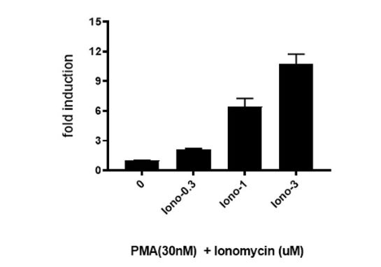
The NFAT Luciferase Reporter CHO cell line (PKC/Ca2+ Signaling Pathway) contains a firefly luciferase gene under the control of NFAT response element stably integrated into CHO K1 cells. This cell line is validated for its response to phorbol 12-myristate 13-acetate (PMA) in the presence of ionomycin.
Interested in screening and profiling inhibitors or activators of NFAT-mediated PKC/Ca2+ pathway signaling without the need to purchase and license the cell line? Check out our Cell Signaling Pathway Screening.
Purchase of this cell line is for research purposes only; commercial use requires a separate license. View the full terms and conditions.
Materials Required for Cell Culture
| Name | Ordering Information |
| Thaw Medium 3 | BPS Bioscience #60186 |
| Growth Medium 3 | BPS Bioscience #79539 |
Materials Required for Cellular Assay
| Name | Ordering Information |
| Assay Medium: Thaw medium 3 | BPS Bioscience #60186 |
| Growth Medium 3D | BPS Bioscience #79539 |
| PMA | LC Laboratories #P1680 |
| Ionomycin | Sigma #l3909 |
| 96-well tissue culture treated white clear-bottom assay plate | Corning #3610 |
| ONE-Step™ Luciferase Assay System | BPS Bioscience #60690 |
| Luminometer |
The cell line has been screened to confirm the absence of Mycoplasma species.
The protein kinase C (PKC)/ Ca2+ response pathway leads to activation of the transcription factor nuclear factor of activator T cells (NFAT). NFAT is regulated by Ca2+ and the Ca2+/calmodulin-dependent serine phosphatase calcineurin. NFAT proteins are phosphorylated and reside in the cytoplasm in resting cells; upon stimulation, they are dephosphorylated by calcineurin, translocate to the nucleus, and induce gene expression.
 Powered by Bioz
San Diego, CA 92121
858-202-1401
Products are for Research Use Only
COMPANY
Copyright © 2024
BPS Bioscience, Inc.
All rights reserved.
Diagrams on this website are created with BioRender.com.