NFAT Reporter (eGFP) Jurkat Recombinant Cell Line
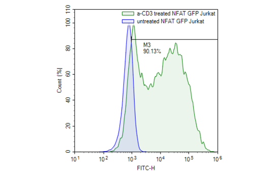
Recombinant Jurkat T-cells expressing an enhanced GFP (eGFP) gene under the control of NFAT response elements located upstream of the minimal TATA promoter. Activation of the NFAT signaling pathway can be monitored by examining eGFP expression.
Interested in screening and profiling inhibitors or activators of NFAT-mediated signaling pathways without the need to purchase and license the cell line? Check out our Cell Signaling Pathway Screening.
Purchase of this cell line is for research purposes only; commercial use requires a separate license. View the full terms and conditions.
Media Required for Cell Culture
| Name | Ordering Information |
| Thaw Medium 2 | BPS Bioscience #60184 |
| Growth Medium 2E | BPS Bioscience #79638 |
The cell line has been screened to confirm the absence of Mycoplasma species.
The Nuclear Factor of Activated T-Cells (NFAT) family of transcription factors plays an important role in mediating the immune response. T-cell activation through the T-cell synapse results in calcium influx. Increased intracellular calcium levels activate the calcium-sensitive phosphatase Calcineurin, which rapidly dephosphorylates the serine-rich region (SRR) and SP-repeats in the amino termini of NFAT proteins. This results in a conformational change that exposes a nuclear localization signal (NLS) promoting NFAT nuclear translocation and inducing gene expression, including various cytokines (IL-2, IL-3, IL4, and TNF-alpha). Members of the NFAT family have been found in many tissue types, including the heart, skeletal muscle and brain. This reporter cell line is designed to monitor T-cell activation or inhibition through various checkpoint inhibitors. It can be used as a control or parental cell line to co-express various immune checkpoint inhibitors, such as PD1.
 Powered by Bioz
San Diego, CA 92121
858-202-1401
Products are for Research Use Only
COMPANY
Copyright © 2024
BPS Bioscience, Inc.
All rights reserved.
Diagrams on this website are created with BioRender.com.