Anti–H3K9me3 polyclonal antibody
Catalog #
25272
$415
*
●
●
Purchase
Description
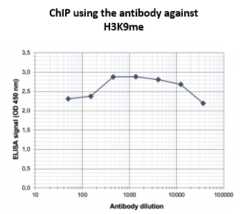
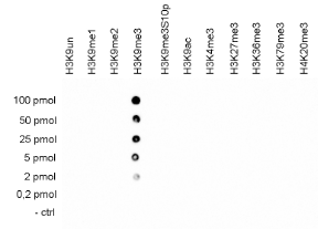
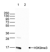
Polyclonal antibody raised in rabbit against the region of histone H3 containing the trimethylated lysine 9 (H3K9me3), using a KLH-conjugated synthetic peptide.
●
Product Data Gallery
Product Info
Storage and Usage
Citations
Clonality
polyclonal
Host Species/Expression System
Rabbit
Purification
Affinity purified
Format
Aqueous buffer solution
Formulation
PBS containing 0.05% azide
Concentration
1.85 mg/ml
Immunogen
synthetic peptide
Background
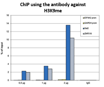
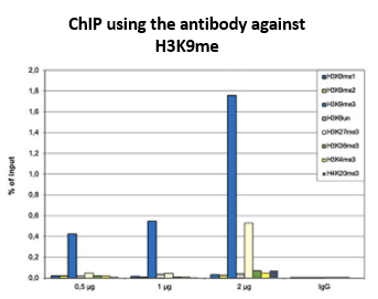
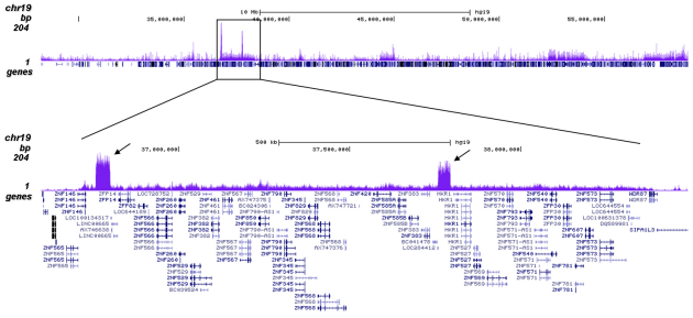
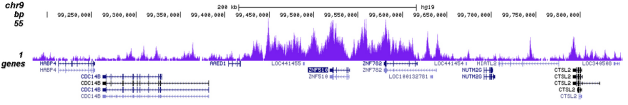
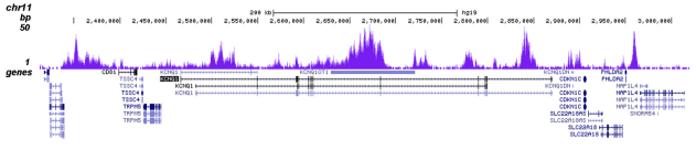
Trimethylation of histone H3K9 is associated with satellite repeat regions and ZNF repeat genes.
 Powered by Bioz
6405 Mira Mesa Blvd. Suite 100
San Diego, CA 92121
858-202-1401
San Diego, CA 92121
858-202-1401
Products are for Research Use Only
COMPANY
Copyright © 2024
BPS Bioscience, Inc.
All rights reserved.
Diagrams on this website are created with BioRender.com.