Notch1dE Lentivirus
Notch1dE Lentiviruses are replication incompetent, HIV-based, VSV-G pseudotyped lentiviral particles that are ready to transduce most mammalian cells, including primary and non-dividing cells. These lentiviruses express a truncated human Notch1 construct (Notch1dE) in which the entire extracellular domain was deleted. The lentiviruses also contain a puromycin selection marker (Figure 1). Once expressed in a cell line of interest, Notch1dE is cleaved by γ-secretase, resulting in the constitutively active intracellular domain of Notch (NICD). NICD translocates to the nucleus, binds to the transcription factor CSL (CBF1/RBPJκ/Suppressor of Hairless/Lag-1) and activates transcription of Notch1-responsive genes.
When used in combination with CSL (CBF1/RBP-Jk) Luciferase Reporter Lentiviruses (BPS Bioscience #78746), NICD activates the CSL (CBF1/RBPJκ/Suppressor of Hairless/Lag-1) responsive elements, inducing luciferase expression.
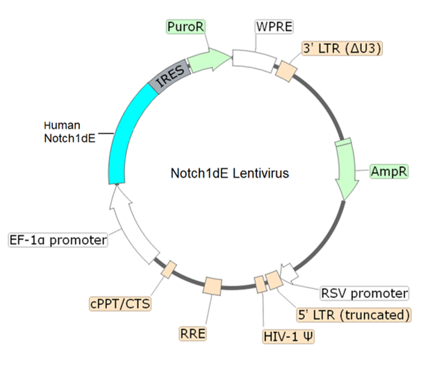
Figure 1. Schematic of the lenti-vector used to generate the Notch1dE lentivirus.

| Name | Ordering Information |
| HEK293 Cells | ATCC #CRL-1573 |
| Thaw Medium 1 | BPS Bioscience #60187 |
| CSL (CBF1/RBP-Jk) Luciferase Reporter Lentivirus | BPS Bioscience #78746 |
| 96-well tissue culture, clear-bottom, white plate | Corning #3610 |
| One-Step™ Luciferase Assay System | BPS Bioscience #60690 |
| Luminometer |
The lentivirus particles were produced in HEK293T cells. They are supplied in cell culture medium containing 90% DMEM + 10% FBS. Virus particles can be packaged in custom formulations by special request, for an additional fee.
The Notch signaling pathway controls cell fate decisions in vertebrates and invertebrates’ tissues and is involved in embryonic development, tissue homeostasis, and regulation of the immune and angiogenic systems. Notch signaling is triggered through the binding of a transmembrane ligand, present in opposing cells, to one of the four existing Notch transmembrane receptors (Notch1/Notch2/Notch3/Notch4). This results in proteolytic cleavage of the Notch receptor, releasing the constitutively active intracellular domain of the Notch receptor (NICD). NICD translocate to the nucleus and associates with the transcription factor CSL (CBF1/RBPJκ/Suppressor of Hairless/Lag-1) and coactivator Mastermind to turn on the transcription of Notch-responsive genes. Dysfunction of Notch signaling has severe consequences, including developmental pathologies or cancer (such as T cell acute lymphoblastic leukemia, T-ALL, and urothelial bladder cancer). The use of Notch inhibitors, mainly gamma-secretase inhibitors, as a cancer therapy option and in the regeneration of tissues is ongoing. Further studies will allow us to have a deeper understanding of Notch signaling and will benefit future therapeutic approaches.
- Lu F.M., et al., 1996 Natl. Acad. Sci. USA 93(11): 5663-5667.
- Kanungo J., et al., 2008 Neurochem. 106: 2236-48.
- Cao L. et al., 2023 Blood Adv. 1182/bloodadvances.2023010380
 Powered by Bioz
San Diego, CA 92121
858-202-1401
Products are for Research Use Only
COMPANY
Copyright © 2024
BPS Bioscience, Inc.
All rights reserved.
Diagrams on this website are created with BioRender.com.