In Vivo-Luc™ Imaging Solution
The In vivo-Luc™ imaging solution is a ready-to-use, sterilized, endotoxin-free solution of D-Luciferin for in vivo imaging of the expression of firefly luciferase. Real-time disease progress or drug efficacy in animals can be monitored in a non-invasive manner using the In vivo-Luc™ imaging solution.
| Catalog # | Name | Amount | Storage |
| 78803-1 | Luciferase Reagent Substrate | 5 ml (5 x 1 ml) | -20°C |
| 78803-2 | Luciferase Reagent Substrate | 25 ml (5 x 5 ml) | -20°C |
15 mg/ml D-Luciferin in Dulbecco's phosphate-buffered saline (DPBS) sterilized and endotoxin free.
Luciferases are enzymes that produce light when they oxidize their substrate. Oxidation of luciferin, the substrate for the most commonly used firefly (Photinus pyralis) luciferase, results in bioluminescence that can be accurately measured using a luminometer and is proportional to the amount of luciferase enzyme present in the system under study. The firefly luciferase reaction requires luciferin, plus adenosine triphosphate (ATP) and Mg2+ as cofactors, and when in the presence of O2, emits a characteristic glow (Figure 1). Common biological experiments that utilize the luciferase reaction are promoter activity assays in which luciferase expression is under the control of a specific promoter, and cell line-derived xenograft (CDX) models in which constitutive luciferase expression allows for real-time, non-invasive monitoring of tumor growth and measurement of disease progression or drug efficacy.
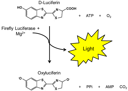
Figure 1. Bioluminescent Reaction Catalyzed by Luciferase.
 Powered by Bioz
San Diego, CA 92121
858-202-1401
Products are for Research Use Only
COMPANY
Copyright © 2024
BPS Bioscience, Inc.
All rights reserved.
Diagrams on this website are created with BioRender.com.