Membrane-Bound TNFα CHO Cell Line
The Membrane-Bound TNFα CHO Cell Line is a clonal CHO cell line stably expressing human membrane-bound TNFα (tumor necrosis factor alpha) driven by an EF1a promoter. The cells were generated by transduction with Membrane-Bound TNFα (mTNFα) Lentivirus (#78955).
Purchase of this cell line is for research purposes only; commercial use requires a separate license. View the full terms and conditions.

Media Required for Cell Culture
| Name | Ordering Information |
| Thaw Medium 3 | BPS Bioscience #60186 |
| Growth Medium 3J | BPS Bioscience #79974 |
Materials Used in Cellular Assay
| Name | Ordering Information |
| Growth Medium 2A | BPS Bioscience #60190 |
| Assay Medium 2A | BPS Bioscience #79621 |
| Thaw Medium 2 | BPS Bioscience #60184 |
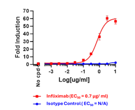
| ADCC Bioassay Effector Cell V Variant (High Affinity)/NFAT Luciferase Reporter Jurkat Cell Line | BPS Bioscience #60541 |
| Infliximab Recombinant Monoclonal Antibody | ThermoFisher #MA5-41776 |
| Clear-bottom, white 96-well tissue culture-treated plate | Corning #3610 |
| ONE-Step™ Luciferase Assay System | BPS Bioscience #60690 |
| Luminometer |
The cell line has been screened to confirm the absence of Mycoplasma species.
Tumor necrosis factor (TNF, also known as TNFα) is produced predominantly by activated macrophages and T lymphocytes. It has been identified as a key regulator in inflammatory and immune responses. TNF signaling pathways are triggered by binding to one of two distinct receptors, designated TNFR1 (TNF receptor 1) and TNFR2, which are differentially regulated on various cell types in normal and diseased tissues. TNFα exists in both a trimeric membrane-bound form (mTNFα) and as a soluble protein. TNFα is synthetized in a precursor form, a cell surface type II transmembrane protein, which is cleaved by metalloproteinases such as TACE (TNFα converting enzyme) into a soluble peptide. Soluble TNFα can then bind to its receptors and activate downstream signaling pathways. Transmembrane TNFα can also bind to TNFα receptors and induce cellular responses. For instance, it enhances cytotoxicity of NK cells, while in the liver it can trigger hepatitis. Anti-TNFα antibodies can bind to the mTNFα and trigger antibody-dependent cell-mediated cytotoxicity (ADCC) or complement-dependent cytotoxicity (CDC) to destroy the mTNFα-expressing inflammatory cells, being a promising therapy for inflammatory diseases.
Wang F., et al., 2017 Mol Med Rep 16: 1021-1030.
Horiuchi T., et al., 2010 Rheumatology 49(7):1215-1228.
 Powered by Bioz
San Diego, CA 92121
858-202-1401
Products are for Research Use Only
COMPANY
Copyright © 2024
BPS Bioscience, Inc.
All rights reserved.
Diagrams on this website are created with BioRender.com.