MART-1 TCR (DMF4) CD8+ NFAT-Luciferase Reporter Jurkat Cell Line
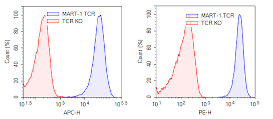
MART-1 TCR (DMF4) CD8+ NFAT-Luciferase Reporter Jurkat Cell Line is a Jurkat cell line generated from the T Cell Receptor (TCR) Knockout NFAT Luciferase Reporter Jurkat Cell Line (BPS Bioscience #78556) by overexpression of human CD8 (NM_001768.6) and MART-1-specific TCR (DMF4) using lentiviral transduction (CD8a Lentivirus #78648 and MART-1-Specific TCR Lentivirus Clone DMF4 #78678). The human TCR clone DMF4 specifically recognizes tumor antigen MART-1 (Melanoma-associated antigen recognized by T cells-1, or Melan-A).
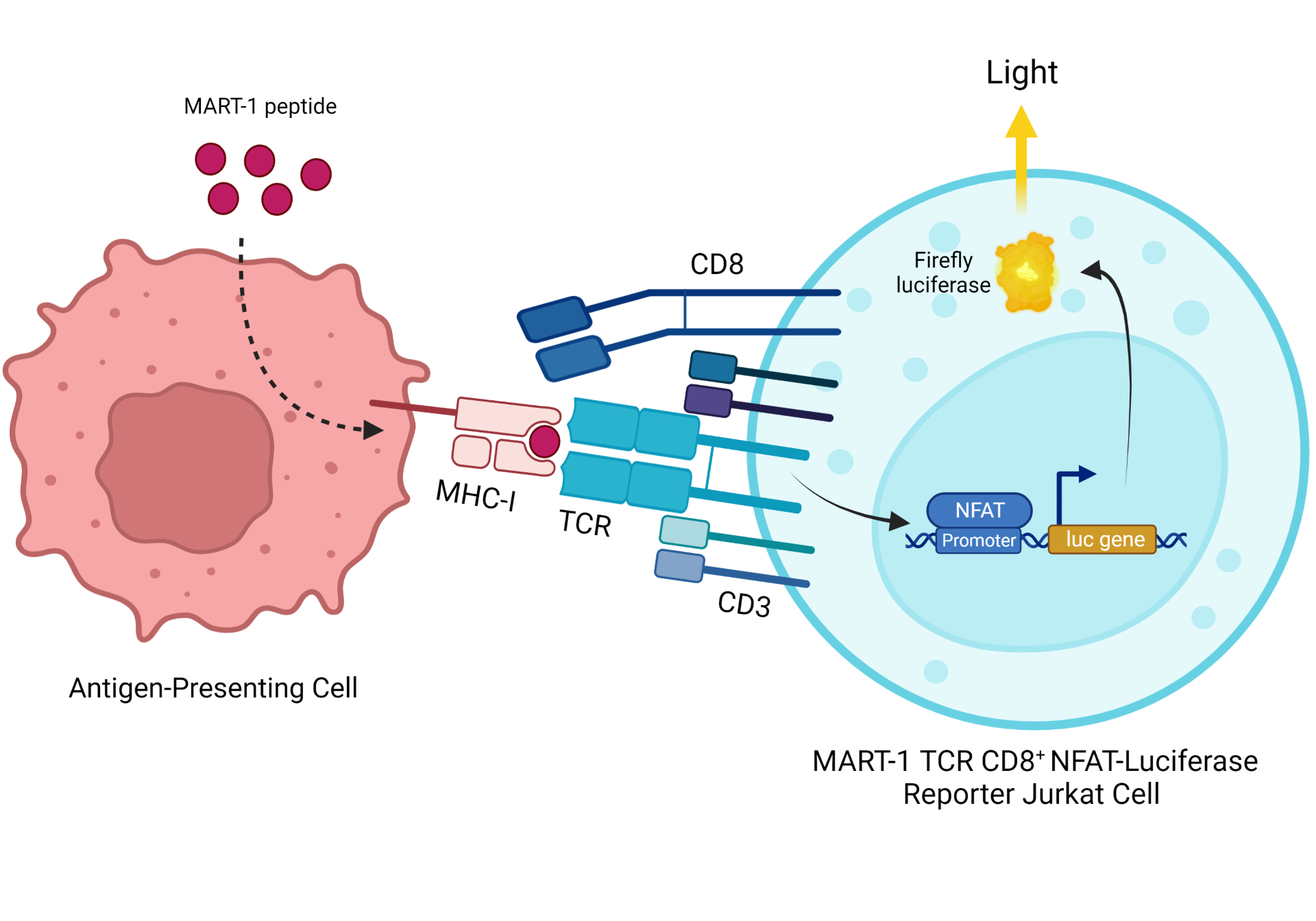
Figure 1: Illustration of the functional co-culture assay used to validate the MART-1 TCR (DMF4) CD8+ NFAT-Luciferase Reporter Jurkat cell line.
Purchase of this cell line is for research purposes only; commercial use requires a separate license. View the full terms and conditions.
| Name | Ordering Information |
| Thaw Medium 2 | BPS Bioscience #60184 |
| Growth Medium 2T | BPS Bioscience #78756 |
| Assay Medium 2D | BPS Bioscience #78755 |
| CD8+ TCR KO NFAT Luciferase Reporter Jurkat Cell Line | BPS Bioscience #78757 |
| T2 Cell Line | ATCC #CRL-1992 |
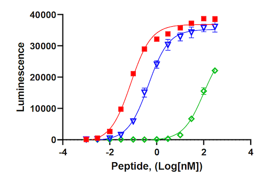
| MART-1 (26-35) Peptide | BPS Bioscience #78759 |
| MART-1 (26-35, Leu27) peptide | BPS Bioscience #78760 |
| MART-1 (27-35) Peptide | BPS Bioscience #78761 |
| APC MHC I Dextramer (HLA-A*02:01 ELAGIGILTV) | Immudex #WB02162 |
| PE anti-human α/β T Cell Receptor Antibody | Biolegend #306707 |
| ONE-Step™ Luciferase Assay System | BPS Bioscience #60690 |
| 96-well tissue culture plate, white, clear bottom |
The cell line has been screened to confirm the absence of Mycoplasma species.
MART-1 is a differentiation antigen expressed on the surface of melanocytes. A peptide fragment of the protein is found in MHC complexes that are recognized by CD8+ cytotoxic T cells. MART-1 is present in most skin cancers, including melanomas, and is used as a biomarker for diagnostic purposes. Since the expression of the protein is restricted to melanocyte containing tissues (skin and retina), and is not found in other tissues, MART-1 is an attractive target for cancer vaccines and adoptive cell therapy. The MART-1 peptide 26-35 is a fragment commonly associated with MHC and recognized by T cell receptors.
CD8 (Cluster of Differentiation 8) is a co-receptor of TCR and a typical marker of cytotoxic T cells. The TCR protein complex is found on the surface of T cells and is responsible for recognizing antigens bound to MHC (Major Histocompatibility Complex) molecules. Stimulation of the TCR results in activation of downstream NFAT (Nuclear factor of Activated T-cells) transcription factors that induce the expression of various cytokines such as interleukin-2 to 4, and TNF-alpha. The use of engineered TCR allows T cells to target specific antigens present in cancer cells, via the MHC, expanding the portfolio of antigens that can be targeted in cancer cell therapy.
 Powered by Bioz
San Diego, CA 92121
858-202-1401
Products are for Research Use Only
COMPANY
Copyright © 2024
BPS Bioscience, Inc.
All rights reserved.
Diagrams on this website are created with BioRender.com.