MAGE-A1-Specific TCR Lentivirus (Clone 1367)
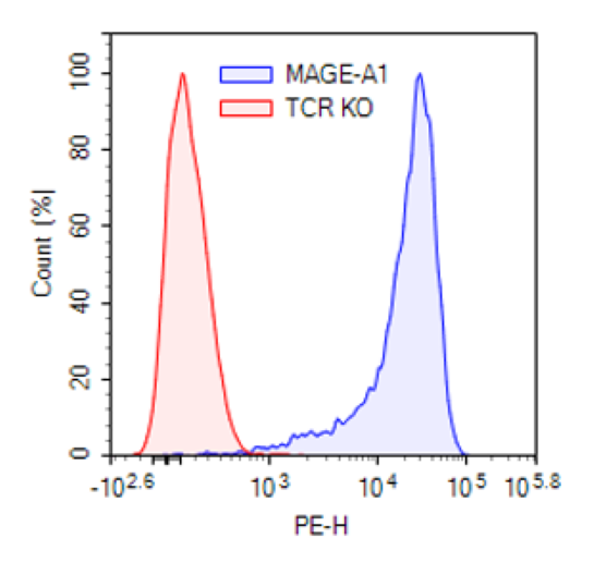
MAGE-A1-Specific TCR Lentivirus (Clone 1367) are replication incompetent, HIV-based, VSV-G-pseudotyped lentiviral particles ready to transduce nearly all types of mammalian cells, including primary and non-dividing cells. These viruses transduce cells with a TCR (T cell receptor) (clone 1367) that specifically recognizes the human antigen MAGE-A1 (Melanoma-associated antigen 1), and in which the TCR α chain and β chain are linked by P2A. The lentiviruses also transduce a puromycin selection marker (Figure 1). Mouse TCR constant regions were used in this construct to boost expression.
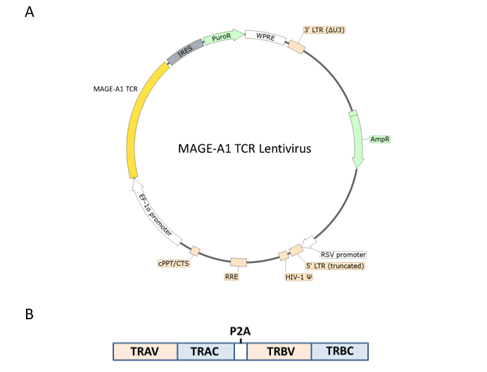
Figure 1. (A) Schematic of the lenti-vector used to generate the MAGE-A1-Specific TCR Lentivirus (Clone 1367) and (B) diagram of the construct, showing the components of the MAGE-1-specific TCR.
TRAV and TRAC correspond to the TCR alpha chain variable and constant regions, respectively, whereas TRBV and TRBC correspond to the TCR beta chain variable and constant regions. Mouse TCR constant regions were used in this construct to boost expression
| Name | Ordering Information |
| Thaw Medium 2 | BPS Bioscience #60184 |
| Growth Medium 2C | BPS Bioscience #79592 |
| Assay Medium 2D | BPS Bioscience #78755 |
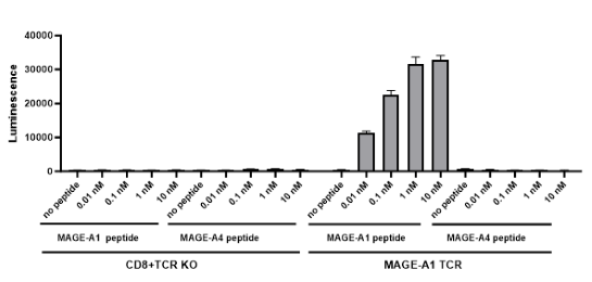
| CD8+ TCR Knockout NFAT-Luciferase Reporter Jurkat Cell Line | BPS Bioscience #78757 |
| T2 Cell Line | ATCC #CRL-1992 |
| MAGE-A1 Peptide (278-286) | BPS Bioscience #78965 |
| MAGE-A4 Peptide (230-239 | BPS Bioscience #78966 |
| PE anti-mouse TCR β chain Antibody | BioLegend #109207 |
| ONE-Step™ Luciferase Assay System | BPS Bioscience #60690 |
| Lenti-Fuse™ Polybrene Viral Transduction Enhancer | BPS Bioscience #78939 |
The lentiviruses were produced in HEK293T cells in medium containing 90% DMEM + 10% FBS. Virus particles can be packaged in custom formulations by special request, for an additional fee.
MAGE (melanoma associated antigen) proteins are CT (cancer testis) antigens, and there are about 60 proteins in the MAGE family that can be subdivided into type I (present only on the X-chromosome, MAGE-A, B and C) and type II (MAGE D-L and necdin). Under normal conditions they are mostly found in the testis and placenta. They are found at high levels in several cancer types, such as melanoma, brain, and breast cancer, and are involved in the development of resistance to chemotherapy, cell motility and cell survival. Expression of MAGE proteins tend to correlate with a poor prognosis. They are intracellular proteins, with MAGE-A1 being mostly cytosolic, making them poor targets for strategies such as CAR-T cell therapy. MAGE proteins are degraded in the proteosome, and the peptides created can then be found on the cell membrane in combination with MHC (major histocompatibility complex) I. The presentation on the cell surface in this form makes them an attractive target for TCR (T cell receptor)-T cell therapy. Several clinical trials are ongoing, and either alone or in combination with other forms of cancer therapy the use of TCR-T cells targeting MAGE-A antigens may become a promising therapeutical avenue.
Matthias O., et al., 2015 Nat Biotechnol 201;33(4):402-7.
Zajac P., et al., 2017 Front. Med. 4:00018.
De Rooji M., et al., 2023 Molecular Therapy Oncology 28:1-14.
 Powered by Bioz
San Diego, CA 92121
858-202-1401
Products are for Research Use Only
COMPANY
Copyright © 2024
BPS Bioscience, Inc.
All rights reserved.
Diagrams on this website are created with BioRender.com.