Cathepsin E Inhibitor Screening Assay Kit
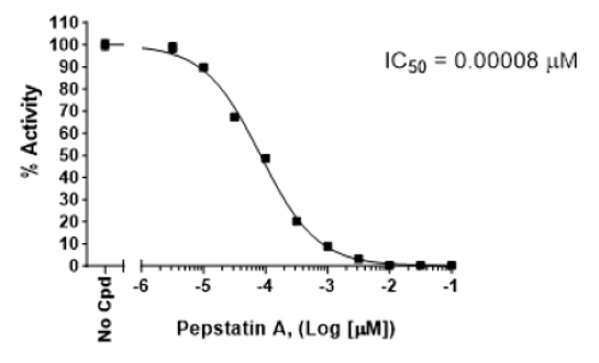
The Cathepsin E Inhibitor Screening Assay Kit is designed to measure the protease activity of Cathepsin E for screening and profiling applications. The Cathepsin E assay kit comes in a convenient 96-well or 384-well format, with enough recombinant human Cathepsin E (amino acids 18-396), its substrate, and Cathepsin buffer for 96 or 384 reactions. This kit includes inhibitor Pepstatin A as control.
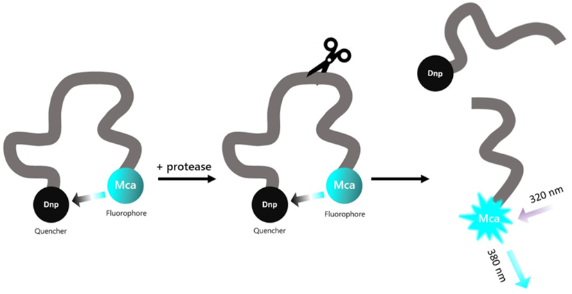
Figure 1: Illustration of the assay principle.
The substrate is an internally quenched fluorogenic substrate. Proteolysis releases the highly fluorescent Mca from the quencher. Fluorescence intensity increases proportionally to the activity of the protease.
Need us to run inhibitor screens or profile your compounds against Cathepsin E? Check out our Protease Screening Services.

- Adjustable micropipettor and sterile tips.
- Fluorescence plate reader capable of measurement at λex330/λem390 nm.
96 reactions
| Catalog # | Name | Amount | Storage |
| 11070 | Cathepsin E, His-Tag* | >1 µg | -80°C |
| CS Substrate 1 | 12.5 µl | -20°C | |
| 4x Cathepsin Buffer | 2 ml | -20°C | |
| 0.5 M DTT | 200 μl | -20°C | |
| 10 mM Pepstatin A | 5 µl | -20°C | |
| 79685 | 384-well black microplate | 1 | Room Temp |
* The concentration of protein is lot-specific and will be indicated on the tube containing the protein.
384 reactions
| Catalog # | Name | Amount | Storage |
| 11070 | Cathepsin E, His-Tag* | >1 µg | -80°C |
| CS Substrate 1 | 2 x 12.5 µl | -20°C | |
| 4x Cathepsin Buffer | 2 x 2 ml | -20°C | |
| 0.5 M DTT | 2x 200 μl | -20°C | |
| 10 mM Pepstatin A | 5 µl | -20°C | |
| 79961 | 384-well black microplate | 1 | Room Temp |
* The concentration of protein is lot-specific and will be indicated on the tube containing the protein.
Cathepsin E, also known as erythrocyte membrane aspartic proteinase, SMP or EMAP, is a homodimer aspartic protease of the peptidase A1 family. It is a non-lysosomal cathepsin, found in the membrane of gastric parietal cells, hepatic cells, proximal tubule in kidney, epithelial cells of the intestine and osteoclasts. It is also found in the endosomes of macrophages, dendritic cells, and microglia. It is involved in antigen processing on the MHC (major histocompatibility complex) class II pathway and age-related neuronal death. It has been linked to gastric and pancreatic cancers such as pancreatic ductal adenocarcinoma (PDAC), and deficiency in cathepsin E plays a role in inflammatory diseases of the skin, such as atopic dermatitis. PDAC is one of the leading causes of death in the context of cancer, mainly due to the lack of early markers of the disease. Cathepsin E is found at high levels in gastric, cervical, esophageal and lung adenocarcinomas, and others, making it an attractive biomarker and imaging peptide probe. Inhibitors that selectively act on cathepsin E and not on cathepsin D have been difficult to identify. Future studies are required to delineate the exact role of cathepsin E in cancer and to develop corresponding therapeutic approaches.
Pontious C., et al., 2019 Pancreatology 19 (7): 951-956.
 Powered by Bioz
San Diego, CA 92121
858-202-1401
Products are for Research Use Only
COMPANY
Copyright © 2024
BPS Bioscience, Inc.
All rights reserved.
Diagrams on this website are created with BioRender.com.