Homogeneous Caspase-7 Assay Kit
Homogeneous Caspase-7 Assay Kit is a kit designed to measure caspase-7 activity for screening and profiling applications. The assay kit comes in a convenient 96-well format, with enough recombinant caspase-7, fluorogenic substrate, and assay buffer for 100 enzyme reactions. This kit also contains Ac-DNLD-CHO, a potent caspase-7 inhibitor, as control.
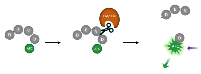
Figure 1: Illustration of the mechanism behind the Homogeneous Caspase-7 Assay Kit.
The substrate Ac-DEVD-AFC is incubated with caspase-7. Proteolysis of the substrate releases the AFC fluorophore. Fluorescence intensity can be measured with a microplate reader able to read fluorescence with λexc=400 nm and λem = 505 nm. Fluorescence is thus proportional to caspase-7 activity.
- Fluorimeter capable of excitation at λ=400 nm and detection at λ=505 nm
- Adjustable micropipettor and sterile tips
- Orbital shaker
| Catalog # | Name | Amount | Storage |
| 70000 | Caspase-7, His-Tag* | 5 µg | -80°C |
| 0.1 mM Caspase-7 Substrate | 50 µl | -80°C | |
| 5x Caspase Assay Buffer 1 | 20 ml | -20°C | |
| 0.5 M DTT | 200 µl | -20°C | |
| 100 µM Ac-DNLD-CHO | 20 µl | -80°C | |
| 79685 | Black, low binding microtiter plate | 1 | Room Temp |
*The concentration of the protein is lot-specific and will be indicated on the tube.
Caspase-7 is a cysteine-aspartic acid protease of the caspase family of proteins, belonging to group II (apoptotic executioner caspases). It plays a role in cell differentiation, tissue regeneration, apoptosis and neural development. It is synthesized as a zymogen, or procaspase, requiring cleavage by initiator caspases to trigger apoptosis. Its activity results in several cell changes, such as membrane blebbing, DNA fragmentation and the formation of apoptotic vesicles. Cell death via apoptosis can occur by intrinsic and extrinsic pathways. In response to DNA damage, metabolic or ER (endoplasmic reticulum) stress the intrinsic pathway can be activated. One example is the use of chemotherapeutic drugs. The release of cytochrome c from the mitochondria results in the formation of the APAF1 (apoptotic protease activating factor 1) apoptosome, recruitment of caspase-9 and finally conversion of procaspase-7 to caspase-7. Cells keep a tight control of caspase activity and express a number of inhibitors such as IAP (inhibitor of apoptosis) proteins. Caspase-7 can be inhibited by XIAP (X-linked IAP), cIAP1 and cIAP2. Deregulation of caspase activity can result in cancer, as many of the regulators of their activity are oncogenes or tumor suppressors. Caspase-7 has been used in the treatment of cancer, osteoarthritis, heart failure and neurogenerative disorders. The use of caspase-3/7 non-specific inhibitor have proved beneficial in maintaining cartilage homeostasis, and potentially preventing and treating osteoarthritis. Further understanding of the role of caspase-7 in disease will allow us to develop innovative and efficacious new therapies.
Chai, J., et al., 2001, Cell 104(5):769-80.
Denault, J.B. and Salvesen, GS., 2003 J. Biol. Chem., 278(36):34042-50.
Boice A. and Bouchier-Hayes L., 2002, Biochimica et Biophysica Acta 1867 (6): 118688.
Lee D., et al., 2000 Molecular Basis of Cell and Developmental Biology 275(21):16007-16014.
 Powered by Bioz
San Diego, CA 92121
858-202-1401
Products are for Research Use Only
COMPANY
Copyright © 2024
BPS Bioscience, Inc.
All rights reserved.
Diagrams on this website are created with BioRender.com.