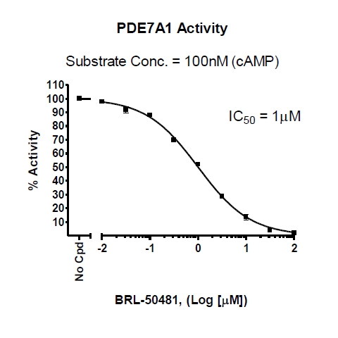
The PDE7A Assay Kit is designed for identification of PDE7A inhibitors using fluorescence polarization. The assay is based on the binding of a fluorescent nucleotide monophosphate generated by PDE7A to the binding agent. PDE7A catalyzes the hydrolysis of the phosphodiester bond in dye-labeled cyclic adenosine monophosphate (cAMP). Nanoparticle beads selectively bind the phosphate group in the nucleotide product. This increases the size of the nucleotide relative to unreacted cAMP. Since the degree of polarization of a fluorophore is inversely related to its molecular rotation, dyes attached to the slowly-rotating nucleotide-bead complexes will not have time to reorient and therefore will emit highly polarized light. Conversely, dyes attached to the rapidly-rotating cyclic monophosphates will obtain random orientations and emit light with low polarization. The key to the PDE7A Assay Kit is the specific binding agent. Using this kit, only two simple steps on a microtiter plate are required for PDE7A reactions. First, the fluorescently labeled cAMP is incubated with a sample containing PDE7A for 1 hour. Second, a binding agent is added to the reaction mix to produce a change in fluorescent polarization. The FP signal is measured using a fluorescent microplate reader capable of measuring fluorescence polarization.
The PDE7A inhibitor screening assay kit comes in a convenient 384-well format, including purified PDE7A enzyme, fluorescently labeled PDE7A substrate (cAMP), binding agent, and PDE assay buffer for 384 enzyme reactions.
Format
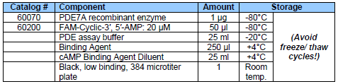
COMPONENTS:
UniProt #
Q13946-2
Background
Phosphodiesterases (PDEs) play an important role in the dynamic regulation
of cAMP and cAMP signaling. PDE7A is widely expressed in various tissues including skeletal
muscle, T lymphocytes, brain and pancreas and plays and an important role in the regulation of
osteoblastic differentiation.
References
1. Malik, R. et al. (2008) Appl. Microbiol. Biotechnol.77 (5): 1167-1173. 2. Pekkinen, M. et al. (2008) Bone. 43 (1): 84-91.
Storage/Stability
Stable at least 6 months from date of receipt, when stored as directed. Kit components require different storage conditions. Be sure to store each component at the proper temperature upon arrival.
Instructions for Use
See assay kit data sheet for detailed protocol
Applications
Useful for studying enzyme kinetics and screening small molecular inhibitors for drug discovery and HTS applications.
Shipping Temperature
-80°C (dry ice)
Warnings
This assay requires a fluorescent microplate reader capable of measuring fluorescence polarization (FP) and equipped with the required partsto read the FP signal. For more information FP technology, visit our Tech Note: FP, assay principles and applications.
Avoid freeze/thaw cycles.
Disclaimers and Limitations
BPS Bioscience assay kits are validated using the listed components according to our specific protocol. Any deviations to kit components or protocols, such as using different proteins, cell/tissue lysates, or buffers (including using the same component from other commercial sources) will void any warranties on the performance of the assay kit and is not recommended.
End-users should immediately report any issues to [email protected] upon receipt of the kit, such as missing components, damaged vials, no dry ice, etc.
 Powered by Bioz