PD-1:PD-L2 Cell-Based Inhibitor Screening Assay Kit
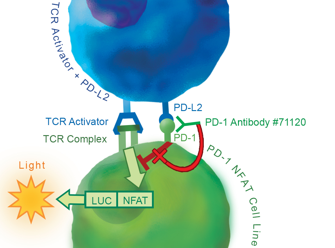
The PD-1/PD-L2 Inhibitor Screening Cell-Based Assay is a bioluminescent cell-based assay that can be used to screen and profile inhibitors of the PD-1:PD-L2 interaction. The assay consists of two main components:
• Growth-Arrested PD-1 Effector cells (PD-1/NFAT reporter-Jurkat cells): Reporter Jurkat T cells expressing firefly luciferase gene under the control of NFAT response elements and also constitutively expressing Human PD-1. These cryopreserved cells are provided in a thaw-anduse format that does not require cell propagation. These cells are modified not to be expanded and are intended to be used in a single experiment.
• Expression vectors for TCR activator and Human PD-L2: Transfection-ready vectors are used to transfect cells to create the target cells that overexpress PD-L2 and an engineered cell surface T cell receptor (TCR) activator.
Purchase of this cell line is for research purposes only; commercial use requires a separate license. View the full terms and conditions.
• HEK293 cell and its growth medium (Thaw medium 1, #60187) or other cell lines
• Transfection reagent for mammalian cell line [We use Lipofectamine™ 2000 (Life technologies #11668027). However, other transfection reagents work equally well.]
• Opti-MEM I Reduced Serum Medium (Life technologies #31985-062)
• 96-well tissue culture-treated white clear-bottom assay plate
• Luminometer
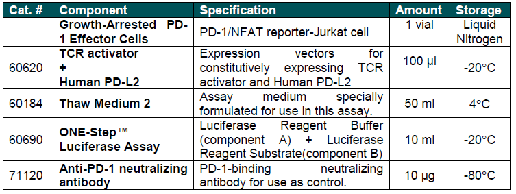
COMPONENTS:
 Powered by Bioz
San Diego, CA 92121
858-202-1401
Products are for Research Use Only
COMPANY
Copyright © 2024
BPS Bioscience, Inc.
All rights reserved.
Diagrams on this website are created with BioRender.com.