PD-1:PD-L1 Cell-Based Inhibitor Screening Assay Kit
The PD-1:PD-L1 Cell-Based Inhibitor Screening Assay is a bioluminescent cell-based assay for screening and profiling inhibitors of the PD-1:PD-L1 interaction. The assay consists of two main components: effector cells and expression vectors encoding TCR activator and human PD-L1. Growth-Arrested PD-1 Effector Cells (PD-1/NFAT Reporter Jurkat cells) are Jurkat cells expressing firefly luciferase, under the control of NFAT response elements, and human PD-1. These cryopreserved cells are provided in a thaw-and-use format that does not require cell propagation. These cells cannot be expanded and are intended to be single use. The expression vectors for TCR Activator and human PD-L1 are transfection-ready vectors that can be used to transfect cells and create target cells that overexpress PD-L1 and an engineered cell surface T cell receptor (TCR) activator. The kit contains enough cells and plasmids for 100 assays if using a 96-well plate.
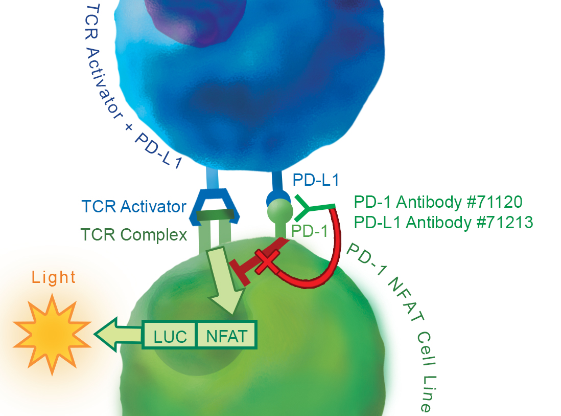
Figure 1: Illustration of the principle of the PD-1:PD-L1 Cell-Based Inhibitor Screening Assay.
Jurkat T cells expressing NFAT reporter with constitutive expression of PD-1 (PD-1/NFAT Reporter Jurkat Cells) act as effector cells. When co-cultivated, TCR complexes on PD-1/NFAT Reporter Jurkat cells are activated by the TCR activator expressed in the transfected cell line of interest, resulting in expression of the NFAT luciferase reporter. However, PD-1 and PD-L1 ligation prevents TCR activation and suppresses the NFAT-responsive luciferase activity. This inhibition can be specifically reversed by anti- PD-1 or anti-PD-L1 antibodies. Neutralizing antibodies block PD-1:PD-L1 interaction and result in reactivation of the NFAT responsive luciferase reporter.
- Cell line of interest, such as HEK293 cells, and corresponding culture media
- Transfection Reagent, such as Lipofectamine™ 2000 Transfection Reagent (Thermo Fisher #11668027)
- Opti-MEM I Reduced Serum Medium (Thermo Fisher #31985-062)
- 96-well tissue culture-treated white clear-bottom assay plate
- Luminometer
| Catalog # | Name | Amount | Storage |
| 79687 | Growth-Arrested PD-1 /NFAT Reporter Jurkat Cell Line | 1 Vial | LN2 |
| 60610 | TCR Activator + Human PD-L1 (expression vectors for expressing TCR Activator and human PD-L1) | 100 µl | -20°C |
| PD-1 Assay Medium | 50 ml | 4°C | |
| 60690 | ONE-Step™ Luciferase Assay | 10 ml | -20°C |
| 71120 | Anti-PD-1 Neutralizing Antibody | 10 µl | -80°C |
The cells have been screened to confirm the absence of Mycoplasma species.
PD-L1 and PD-L2 binding to PD-1, a receptor expressed on T-cells, negatively regulates immune responses. PD-1 ligands PD-L1 and PD-L2 are found on the surface of many cancer cells, and their interaction with receptor PD-1 inhibits T cell activity and allows cancer cells to escape immune surveillance. This pathway is also involved in regulating autoimmune responses. Therefore, these proteins (termed immune checkpoints) are promising therapeutic targets for many types of cancer as well as multiple sclerosis, arthritis, lupus, and type I diabetes. Checkpoint inhibitors have remarkable efficacy in a wide range of cancer types and have revolutionized cancer treatment. PD-1 inhibitors nivolumab, pembrolizumab, cemiplimab and PD-L1 inhibitors atezolizumab, avelumab, and durvalumab are all FDA-approved drugs for immuno-therapy.
Sasca D, et al. (2019) Blood 133: 2305-2319.
 Powered by Bioz
San Diego, CA 92121
858-202-1401
Products are for Research Use Only
COMPANY
Copyright © 2024
BPS Bioscience, Inc.
All rights reserved.
Diagrams on this website are created with BioRender.com.