NFAT Luciferase Reporter Jurkat Cell Line
The NFAT Luciferase Reporter Jurkat Cell Line expresses firefly luciferase under the control of the NFAT response elements stably integrated into the Jurkat cell genome. This reporter cell line is designed to monitor T cell activation as well as inhibition by various immune checkpoint inhibitors; it can be used as a control or parental cell line to co-express various immune checkpoint inhibitors, such as PD-1.
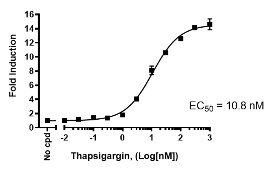
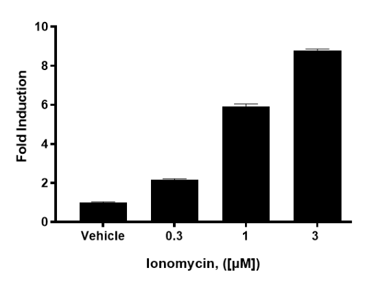
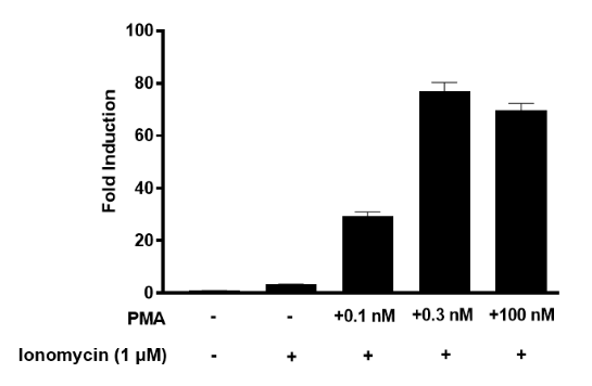
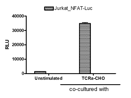
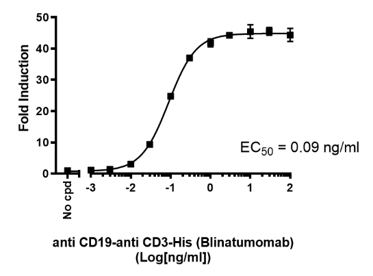
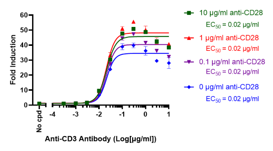
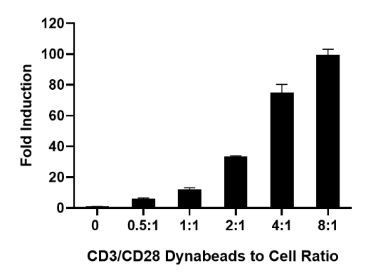
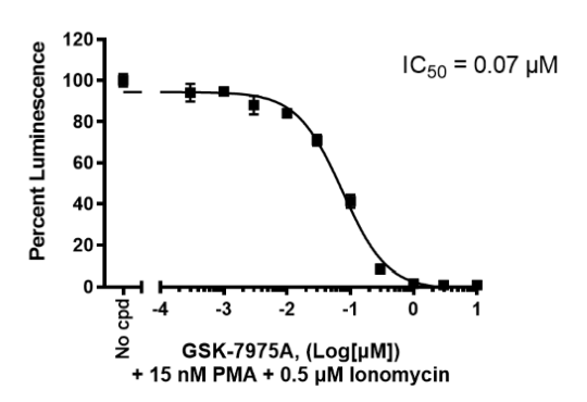
This cell line has been functionally validated and responds to thapsigargin, ionomycin, phorbol 12-myristate 13-acetate (PMA), anti-CD3 antibodies, and Dynabeads™ Human T-Activator CD3/CD28. It can be used to measure T cell activation through a variety of TCR activators including TCR activator (anti-CD3ε scFv)/CHO cells (BPS Bioscience #60539) and CD3 x CD19 bispecific antibody (Blinatumomab) in the presence of CD19+ Raji cells.
Interested in screening and profiling inhibitors, blocking antibodies, or activators of T cell activation pathways without the need to purchase and license the cell line? Check out our Cell Signaling Pathway Screening.
This product has been cited 17 times.
Purchase of this cell line is for research purposes only; commercial use requires a separate license. View the full terms and conditions.
Media Required for Cell Culture
| Name | Ordering Information |
| Thaw Medium 2 | BPS Bioscience #60184 |
| Growth Medium 2B | BPS Bioscience #79530 |
Materials Required for Cellular Assays Described in the Functional Validation section
| Name | Ordering Infomation |
| Ionomycin | Sigma #I3909 |
| Phorbol 12-myristate 13-acetate (PMA) | LC Laboratories #P1680 |
| TCR Activator CHO Recombinant Cell Line | BPS Bioscience #60539 |
| Thapsigargin | Sigma #T9033 |
| Anti-CD19 x anti-CD3 bispecific antibody | BPS Bioscience #100441 |
| Anti-CD3 antibody | BPS Bioscience #71274 |
| Anti-CD28 antibody | BPS Bioscience #100182 |
| Dynabeads™ Human T-Activator CD3/CD28 | Thermo Fisher #11131D |
| GSK-7975A | Sigma #5.34351 |
| ONE-Step™ Luciferase Assay System | BPS Bioscience #60690 |
| White, clear-bottom cell culture plate, 96-well | |
| Luminometer |
The cell line has been screened to confirm the absence of Mycoplasma species.
Nuclear Factor of Activated T cells (NFAT) is a family of 5 transcription factors of near ubiquitous expression, known to have a central function in the immune system, for example by inducing the expression of various cytokines (such as IL-2, IL-3, IL-4, and TNFα) in T cells. NFATs cooperate with multiple other proteins to regulate distinct gene expression programs that determine the fate and function of T cell populations. The NFAT family also plays important roles in the nervous system, in the heart, and in skeletal muscles.
In resting T cells, the NFAT protein is phosphorylated and confined to the cytoplasm in an inactive state. In response to a stimulus, an influx of calcium activates the Ca2+/calmodulin-dependent serine phosphatase calcineurin, which rapidly dephosphorylates the serine-rich region (SRR) and SP-repeats in the amino termini of NFAT proteins. This results in a conformational change that exposes a nuclear localization signal, promoting NFAT translocation to the nucleus. In the nucleus, NFAT proteins cooperate with other transcriptional regulators to induce gene expression.
Through their role in the immune system NFATs are involved in inflammation and these transcription factors are considered promising therapeutic targets in a variety of immune-related diseases.
1. Clipstone NA, Crabtree GR. Nature. 1992 Jun 25;357(6380):695-7.
2. Lyakh, L., et al. Mol Cell Biol. 1997 May;17(5):2475-84.
 Powered by Bioz
San Diego, CA 92121
858-202-1401
Products are for Research Use Only
COMPANY
Copyright © 2024
BPS Bioscience, Inc.
All rights reserved.
Diagrams on this website are created with BioRender.com.