CD40:CD40L TR-FRET Assay
The CD40:CD40L TR-FRET Assay Kit is designed to measure binding activity of CD40 (cluster of differentiation 40) to CD40L (CD40 ligand) for screening and profiling applications using TR-FRET (Time-Resolved Fluorescence Resonance Energy Transfer). It utilizes Terbium-labeled donor and a labeled acceptor to complete the TR-FRET pairing. The CD40:CD40L TR-FRET Assay Kit comes in a convenient 384-well format, with enough recombinant biotinylated CD40 (amino acids 21-193), recombinant CD40L (amino acids 116-261), Tb-Labeled Donor and Dye-Labeled Acceptor and assay buffer for 384 reactions.
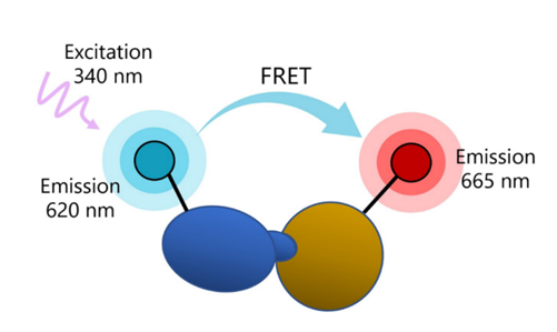
Figure 1: CD40:CD40L TR-FRET Assay Kit schematic.
A sample containing terbium-labeled donor, dye-labeled acceptor, CD40, CD40L, and an inhibitor is incubated for 90 minutes. The fluorescence intensity is then measured using a fluorescence reader. In the presence of binding of CD40 to CD40L, energy transfer occurs due to the proximity of the donor and acceptor. Disruption of the binding results in decrease of energy transfer. Fluorescence intensity at λ=665 nm corresponds directly to the binding of CD40 to CD40L.
Need us to run inhibitor screens or profile your compounds against CD40:CD40L? Check out our Immunotherapy Biochemical Screening Services.
- Fluorescent microplate reader capable of measuring Time Resolved Fluorescence Resonance Energy Transfer (TR-FRET)
- Adjustable micropipettor and sterile tips
| Catalog # | Name | Amount | Storage |
| 79102 | CD40, Fc Fusion (IgG1) Avi-Tag, Biotin Labeled* | 15 µg | -80°C |
| 71191 | CD40L (CD154), His-Tag (Human) | 2 µg | -80°C |
| 30017 | Anti-His Tb-Labeled Donor | 2 x 10 µl | -20°C |
| Dye-Labeled Acceptor | 2 x 10 µl | -20°C | |
| 79311 | 3x Immuno Buffer 1 | 4 ml | -20°C |
| 79969 | White, non-binding Corning low volume microtiter plate | 1 | Room Temp |
* The concentration of protein is lot-specific and will be indicated on the tube containing the protein.
CD40 (cluster of differentiation 40), also known as TNFRSF5 ((tumor necrosis factor)-receptor superfamily 5) is a type I transmembrane protein involved in the activation of antigen presenting cells (APCs), such as dendritic cells, B cells and macrophages. It can also be found in epithelial and endothelial cells, and tumor cells. CD40L (CD40 ligand), also known as TNFSF5 and CD154, is a type II membrane glycoprotein that exists in cells in membrane bound (mCD40L) and soluble (sCD40L) forms. It is found at high levels in activated CD4+ T cells, and at lower levels in Th1, Th2, Th17 and Tregs. Expression can also be induced in NK cells, CD8+ T cells, basophils, and others. CD40 and CD40L are stimulatory immune checkpoints, and their signaling is mediated by different TRAF (TNF receptor associated factor), in a cell and stimuli dependent mode. For example, it mediates the activation of the NF-κB (nuclear factor kappa-B) pathway. CD40 is implicated in Hyper Ig-M immunodeficiency, where IgM is found at high levels in the serum, while other IgG are present at lower-than-normal levels, and patients have higher risk of developing autoimmune diseases and cancer. The role of CD40 and CD40L as immune checkpoints makes them highly attractive targets in cancer therapy, and several clinical trials using anti-CD40 or anti-CD40L agonist antibodies or increasing their expression are underway targeting both hematological and solid tumors. The inhibition of CD40:CD40L interaction is also clinically relevant, and clinical trials have been focusing on treatment options for lupus, rheumatoid arthritis and ALS (amyotrophic lateral sclerosis). Further studies and development of refined therapies will continue to benefit the cancer therapy field and patients suffering from autoimmune disorders.
Annis A., et al., 2004 J. Amer. Chem. Soc. 126(4): 15495-15503.
Yan, T., et al., 2001 J. Cellular Biochem. 83(2): 320-325.
Tang T., et al., 2021 Pharmacol The 219:107709.
 Powered by Bioz
San Diego, CA 92121
858-202-1401
Products are for Research Use Only
COMPANY
Copyright © 2024
BPS Bioscience, Inc.
All rights reserved.
Diagrams on this website are created with BioRender.com.