IKCA1 (KCNN4) HEK293 Cell Line
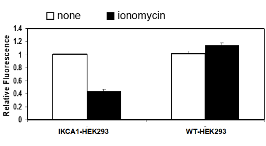
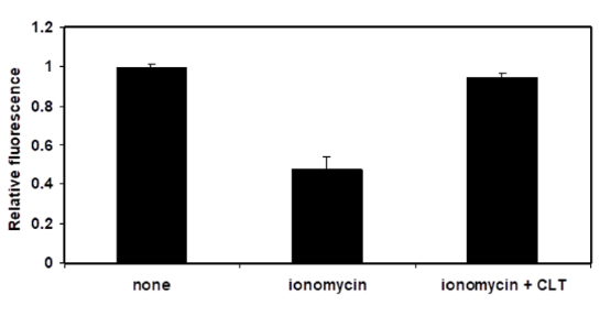
IKCA1 (KCNN4) HEK293 Cell Line are engineered HEK293 cells expressing human IKCA1, also known as KCNN4 (Intermediate conductance calcium-activated potassium channel protein 4), IK1, hKCa4, and hSK4), Genbank Accession No. NM_002250. This cell line has been validated in a cellular assay using a membrane potential-sensitive fluorescence dye (DiBAC4(3)) and by Western blot for IKCA1 expression.
Purchase of this cell line is for research purposes only; commercial use requires a separate license. View the full terms and conditions.
Media Required for Cell Culture
| Name | Ordering Information |
| Thaw Medium 1 | BPS Bioscience #60187 |
| Growth Medium 1B | BPS Bioscience #79531 |
The cell line has been screened to confirm the absence of Mycoplasma species.
IKCA1, also known as Intermediate conductance calcium-activated potassium channel protein 4), IK1, hKCa4, and hSK4, are part of a potentially heterotetrametric voltage-independent potassium channel that is activated by intracellular calcium. Activation is followed by membrane hyperpolarization, which promotes calcium influx. The encoded protein may be part of the predominant calcium-activated potassium channel in T-lymphocytes, but it is also present in endothelial cells and cardiac fibroblasts. They contribute to vascular smooth muscle cell proliferation, migration and cardiac fibrosis. IKCA1 was found to be present at high levels in breast cancer (BC) cells, and when depleted lead to decreased tumorigenesis. IKCA1 can be modulated with small molecules, and these may provide a therapeutical opportunity in the treatment of BC and cardiovascular diseases.
Ghanshani S., et al., J Biol Chem. 275(47): 37137-37149.
Hoffman J.F., et al., 2003 PNAS 100 (12): 7366-7371.
de-Allie F.A., et al., 1996, Br J Pharmacol. 117(3):479-487.
Terstappen G.C., et al., 2003 Neurosci Lett. 346(1-2):85-88.
Gross D., et al., 2022 Cell Death and Disease 13:902.
 Powered by Bioz
San Diego, CA 92121
858-202-1401
Products are for Research Use Only
COMPANY
Copyright © 2024
BPS Bioscience, Inc.
All rights reserved.
Diagrams on this website are created with BioRender.com.