HDAC11 Fluorogenic Assay Kit
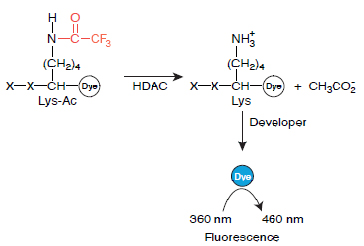
The HDAC11 Fluorogenic Assay Kit is a complete assay system designed to measure histone deacetylase 11 (HDAC11) activity for screening and profiling applications. The HDAC11 Fluorogenic Assay Kit is based on a unique fluorogenic substrate and developer combination. This assay method eliminates dealing with the radioactivity, extraction, and chromatography aspects of traditional assays. Using this kit, only two simple steps on a microtiter plate are needed to analyze the HDAC11 activity level. First, the HDAC fluorometric substrate, containing an acetylated lysine side chain, is incubated with purified HDAC11. The deacetylation sensitizes the substrate so subsequent treatment with the Lysine Developer produces a fluorophore that can then be measured using a fluorescence reader.
HDACs regulate cellular processes by catalyzing the hydrolysis of an acetyl group from acetyllysines in modified proteins. In the HDAC assay, fluorescent-dye molecules are attached to a peptide containing acetyllysine. Attachment to the peptide quenches the fluorescence of the dye. After treatment of the peptide with an HDAC, the reaction is mixed with a development solution that is specific for nonacetylated lysines. If the acetyl group has been removed from the lysine by the HDAC, this solution will release the dye allowing for fluorescence. The amount of fluorescence is therefore directly related to the level of HDAC activity.
Need us to run inhibitor screens or profile your compounds against HDAC11? Check out our Deacetylase/Sirtuin Screening Services.
This product has been cited 4 times.
| Catalog # | Component | Amount | Storage | |
| 50021 | HDAC11 human recombinant enzyme | 10 µg | -80°C | Avoid freeze/ thaw cycles! |
| 50040 | Fluorogenic HDAC substrate class 2A (5 mM) | 25 µl | -80°C | |
| 50030 | 2x HDAC Developer (contains Trichostatin A) (2 µM) |
6 ml | -80°C | |
| Trichostatin A (1 mM) in DMSO | 100 µl | -20°C | ||
| 79320 | HDAC11 Assay Buffer | 10 ml | -20°C | |
| 79685 | black, low binding NUNC black microtiter plate | 1 plate | Room Temp | |
Deubzer, H.E., et al., Int. J. Cancer. 2013 May 1; 132(9):2200-8.
 Powered by Bioz
San Diego, CA 92121
858-202-1401
Products are for Research Use Only
COMPANY
Copyright © 2024
BPS Bioscience, Inc.
All rights reserved.
Diagrams on this website are created with BioRender.com.